


Cancer. Obesity. Heart disease. Alzheimer’s. Parkinson’s. Cerebral palsy. Depression. Some of medicine’s most devastating conditions still have more questions than answers. Armed with innovative resources and bold ambition, WashU Medicine researchers are on a mission to change that.
Activating brown fat could make your metabolism work smarter, not harder
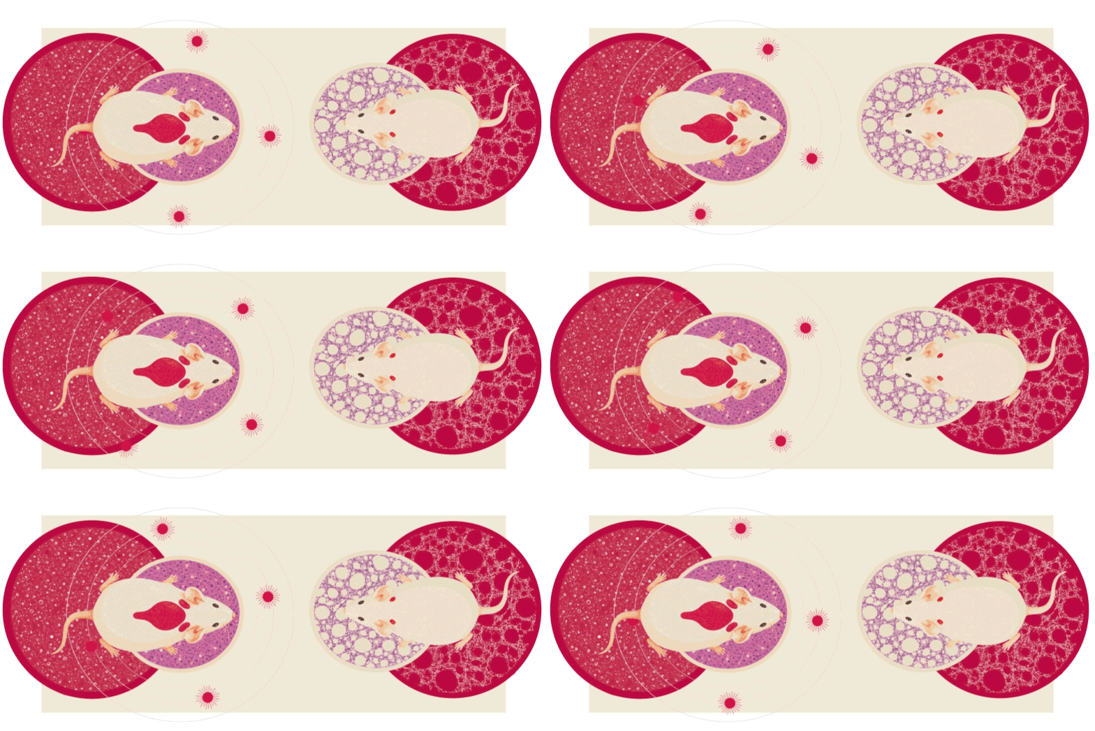
It turns out not all fat is created equal — a discovery in how brown fat, or “good” fat, burns calories could open new doors for supporting weight loss and treating metabolic diseases such as obesity, diabetes and insulin resistance.
“The pathway we’ve identified could provide opportunities to target the energy expenditure side of the weight loss equation, potentially making it easier for the body to burn more energy by helping brown fat produce more heat,” said Irfan Lodhi, PhD, a professor of medicine in the Division of Endocrinology, Metabolism and Lipid Research, who led the study published in Nature. “Boosting this kind of metabolic process could support weight loss or weight control in a way that is perhaps easier to maintain over time than traditional dieting and exercise.”
Scientists have long known that mitochondria — the cell’s main energy producers — in brown fat produce heat. Studying mice, Lodhi and his team uncovered a backup system in which cellular compartments called peroxisomes generate heat involving the protein ACOX2. When exposed to cold, peroxisomes in brown fat increase in number, an effect even more dramatic when the main mitochondrial heat protein is missing, offering strong evidence that peroxisomes act as a compensatory heater.
Lodhi and his colleagues discovered that mice with high levels of ACOX2 in their brown fat showed better weight control and blood sugar regulation, even on unhealthy diets.
The fatty acids processed by ACOX2 occur naturally and are found in dairy and certain gut bacteria, suggesting that dietary interventions such as specific foods or probiotics could boost this heat production pathway, helping people lose weight and improve their metabolic health.
Lodhi and his colleagues are now investigating possible drug compounds that could activate the ACOX2 pathway, moving their discovery closer to clinical applications — and closer to transforming this “good” fat into a powerful tool for fighting metabolic diseases.
Rewiring hope: A new era of precision treatment for Parkinson’s disease
For the more than 10 million people worldwide affected by Parkinson’s disease, reminders of the devastating disorder are everywhere: hand tremors make getting dressed a daily challenge, muscle stiffness leads to chronic pain with every step, restless sleep causes intense exhaustion, and memory and mood changes make previously enjoyable activities undesirable.
For too long, patients and their families have had only one option: mitigate the symptoms. But the disease ravages on, eventually taking over body, mind and spirit. Until now.
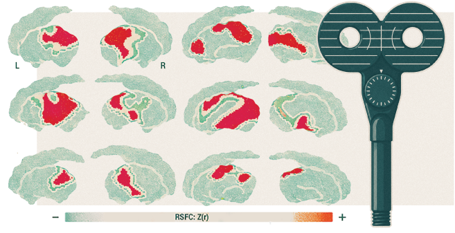
Scientists from WashU Medicine and Changping Laboratory in China have identified a brain network that may be the “command center” for Parkinson’s disease — paving the way for personalized, non-invasive treatment that could slow or even reverse the progression of the disease.
For decades, doctors focused on the basal ganglia, the part of the brain that controls muscle movements, as the source of Parkinson’s. But this didn’t explain why patients also deal with “non-motor” issues, like sleep, memory and digestive problems.
Nico U. Dosenbach, MD, PhD, the David M. & Tracy S. Holtzman Professor of Neurology; Hesheng Liu, PhD, at Changping Laboratory and their respective teams identified the Somato-Cognitive Action Network (SCAN) as the core network responsible for Parkinson’s. First described by Dosenbach in 2023, the SCAN links the parts of the brain responsible for thinking and planning with the parts that control movement. In people with Parkinson’s, this network is “hyperconnected,” disrupting not only movement but also cognitive and bodily functions.
In a clinical trial, 18 patients who received an experimental noninvasive neuromodulation therapy called transcranial magnetic stimulation (TMS) targeting the SCAN more than doubled symptom improvement compared to patients receiving TMS directed at surrounding brain areas.
“This work demonstrates that Parkinson’s is a SCAN disorder,” said Dosenbach. “Changing the activity within SCAN could slow or reverse the progression of the disease, not just treat the symptoms.”
A small implant is delivering big relief for severe depression
What if relief from severe depression didn’t come from a new drug — but from an electrical signal inside our bodies?
In the RECOVER study — a multicenter clinical trial led by WashU Medicine researchers — scientists found that stimulating the vagus nerve improved symptoms and quality of life in patients with advanced treatment-resistant depression. These patients had tried an average of 13 treatments without success and had lived with depression for nearly three decades.
“There is a dire need to find effective treatments for these patients, who often have no other options,” said lead investigator Charles Conway, MD, a professor of psychiatry and director of the WashU Medicine Treatment Resistant Mood Disorders Center. “With this kind of chronic, disabling illness, even a partial response to treatment is life altering, and with vagus nerve stimulation we’re seeing that benefit is lasting.”
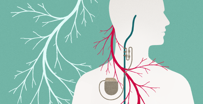
The vagus nerve is one of the body’s main communication lines, carrying signals between the brain and key organs that help regulate mood, heart rate and other physical functions.
Unlike “shock therapy” which delivers brief, high-intensity stimulation to the brain, vagus nerve stimulation (VNS) works more like a pacemaker, sending ongoing signals through that nerve.
The implanted device sits under the skin in the chest and works quietly in the background. This steady approach is key, as the benefits to VNS build over time — nearly one-third of patients who had not responded after one year reported benefits by the end of the second.
“Seeing results for this complicated illness makes me optimistic about the future of this treatment,” said Conway. “These results are highly atypical, as most studies of markedly treatment-resistant depression have very poor sustainability of benefit, certainly not at two years. We’re seeing people getting better and staying better.”
Stopping plaque in its tracks — a new approach to heart disease
Heart disease remains the world’s leading cause of death among adults, contributing to nearly 20 million deaths each year worldwide. And while treatments have long focused on cholesterol as the culprit, a study led by Kory J. Lavine, MD, PhD, may have identified a new target.
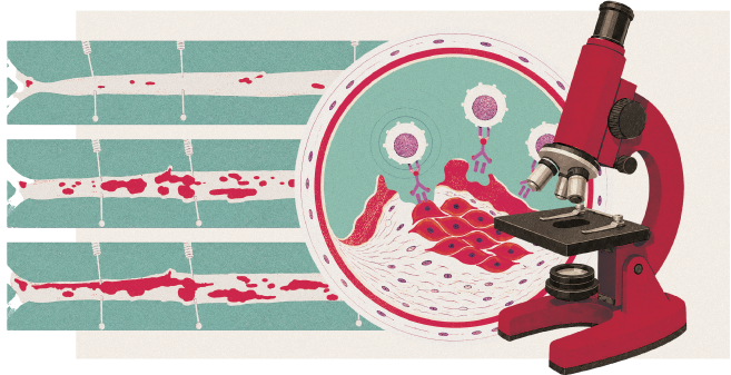
Lavine, who also directs the WashU Medicine Center for Cardiovascular Research, and a team of researchers found that eliminating modulated smooth muscle cells reduced plaque buildup in the arteries of mice — pointing to a new way of treating heart disease beyond a focus on cholesterol.
The strategy uses a form of immunotherapy, a technique already transforming cancer care, to target those cells that contribute to inflammation inside plaques. By disrupting that cycle, researchers were able to reduce plaque size and instability.
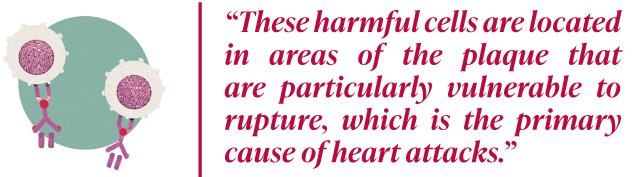
“We found that these harmful cells are located in areas of the plaque that are particularly vulnerable to rupture, which is the primary cause of heart attacks,” said Lavine.
If similar results can be achieved in people, this approach may open the door to new treatments for heart disease, including for patients who don’t fully benefit from existing therapies.
For the millions of people at risk of heart attack or stroke, it could mean a more precise way to stop the deadliest plaques before they do the most harm.
Big steps forward for cerebral palsy
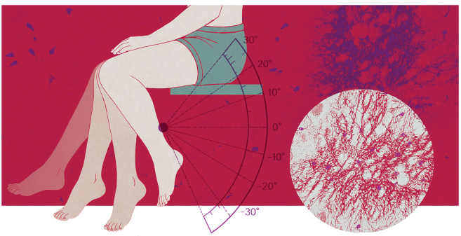
For many children with cerebral palsy, leg dystonia can make walking difficult and even painful. However, a quick test developed by physician-scientists in the Department of Neurology offers a clearer way to assess dystonia, with the potential to guide more precise care.
More than half of children with cerebral palsy experience these involuntary, often painful muscle contractions that lead to abnormal movement and posture. Yet it has long been difficult to diagnose — and even harder to define.
For decades, diagnosis has relied largely on an ultimately inconsistent benchmark: the physician’s subjective assessment.
“An overarching goal of this work is about standardizing dystonia assessment by quantifying diagnoses that were previously based on doctors’ gut feelings,” said Bhooma Aravamuthan, MD, DPhil, an assistant professor of neurology. “These results can be immediately put into clinical practice to help guide treatment.”
In a study led by Aravamuthan, children were recorded performing a simple seated task, allowing specialists to track how their legs moved toward the body’s midline, a subtle but reliable marker of dystonia severity.
The team then turned to mice, stimulating specific brain cells involved in motor control to recreate the same movement pattern, offering clues to the underlying biology and a potential target for future treatments.
What they found was a link between the dystonia-like movement patterns and sustained activity in these brain cells.
This objective approach to evaluating dystonia could be used in clinics right away. Now, the test is already helping guide treatment decisions and allowing doctors to track changes in care with more consistency and confidence.
For families navigating cerebral palsy, this new approach to patient care offers something simple but powerful — actual answers for their child.
Beating the biological clock of Alzheimer’s
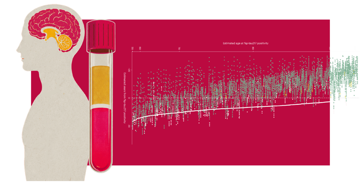
Alzheimer’s disease starts silently — 20 years or more before memory loss and other symptoms ever appear. But what if you could estimate when those symptoms might begin? Thanks to a simple blood test and a mathematical model, researchers have laid the groundwork for doing so.
“Our work shows the feasibility of using blood tests, which are substantially cheaper and more accessible than brain imaging scans or spinal fluid tests, for predicting the onset of Alzheimer’s symptoms,” said senior author Suzanne E. Schindler, MD, PhD, an associate professor in the Department of Neurology. “In the near term, these models will accelerate our research and clinical trials. Eventually, the goal is to be able to tell individual patients when they are likely to develop symptoms, which will help them and their doctors to develop a plan to prevent or slow symptoms.”
Schindler and Kellen K. Petersen, PhD, a mathematician and instructor in neurology, leveraged prior work at WashU Medicine showing that amyloid and tau — the hallmark proteins of Alzheimer’s — accumulate in the brain in a predictable, consistent pattern over decades. Much like tree rings indicate age, Schindler and Petersen found that this protein buildup can be used as a “clock.”
By measuring blood levels of p-tau217, which reflect both amyloid and tau, scientists can determine a person’s position on that clock and estimate their risk of developing Alzheimer’s symptoms. For most people who developed symptoms, the clock also estimated the age that symptoms began within 3-4 years. While this model cannot yet be used on an individual basis, it can “make clinical trials more efficient by identifying individuals who are likely to develop symptoms within a certain period of time,” Petersen said.
This discovery may transform Alzheimer’s from a “wait and see” diagnosis into a timeline, opening the door for scientists to test preventive therapies before brain damage becomes irreversible — and ultimately offering a path to end this devastating disease for families around the world.
A new vaccine fights one of cancer’s deadliest tumors
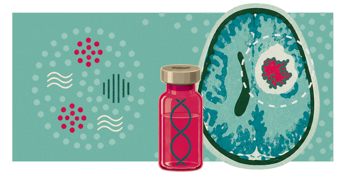
Glioblastoma is one of the deadliest brain cancers. It grows quickly, resists treatment and leaves patients and families facing a devastating truth: even the best available standard of care is rarely enough to stop it.
WashU Medicine physician-scientists refused to accept those odds. Their research led to the development of a personalized vaccine designed to help the immune system recognize and attack each patient’s tumor.
A phase 1 trial involved patients with an unmethylated MGMT subtype of glioblastoma, an especially aggressive form of the cancer. Conducted at Siteman Cancer Center, based at Barnes-Jewish Hospital and WashU Medicine, the trial found that the vaccine caused no serious side effects and prolonged patients’ overall survival compared with historical outcomes after surgery and chemo-radiotherapy.
The study was led by Tanner M. Johanns, MD, PhD, an assistant professor in the Division of Oncology, who treats patients at Siteman and is a research member there.
“This kind of vaccine is a first for glioblastoma,” said Johanns.
In the trial, two-thirds of the patients had no progression six months after surgery, and two-thirds survived one year. Historically, only about 40% of glioblastoma patients reach either benchmark. One-third of participants were still alive after two years, including one patient who remains alive and recurrence-free nearly five years after diagnosis — an extraordinary milestone.
Because glioblastoma is especially difficult for the immune system to recognize and stop, the vaccine is designed around what makes each patient’s tumor unique. Using an algorithm developed at WashU Medicine, researchers identified distinguishing proteins found on a patient’s cancer cells, then created a DNA-based vaccine to help the immune system recognize those telltale proteins and attack the tumor.
The next step for researchers is to assess the vaccine’s efficacy in a larger group of patients and expand treatment to all types of glioblastomas.
The work reflects what scientific research is built to do: move bold ideas from the lab toward the people who can’t wait a second longer.
Published in the Autumn 2026 issue